A diagnostic imaging technique based on the phenomenon of nuclear magnetic resonance (NMR). NMR is a process in which protons interact with a strong magnetic field and with radio waves to generate electrical pulses that can be processed in a similar way to computerized tomography. The medical application of NMR, began in the 1950s, but the first images of live patients were not produced until the late 1970s. Images produced by MRI are similar to those produced by computerized tomography using X-rays, but without the radiation hazard.
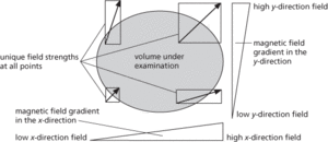
A major factor in the high costs of MRI is the need for a superconducting magnet to produce the very strong magnetic fields (0.1−2 tesla). A niobium–titanium alloy, which becomes superconducting at −269°C, is used to construct the field coils. These need to be immersed in liquid helium. Superimposed on this large magnetic field are smaller fields, with known gradients in two directions. These gradient fields produce a unique value of the magnetic field strength at each point within the instrument.
Some nuclei in the atoms of a patient’s tissues have a spin, which makes them behave as tiny nuclear magnets. The purpose of the large magnetic field is to align these nuclear magnets. Having achieved this alignment, the area under examination is subjected to pulses of radio frequency (RF) radiation. At a resonant frequency of RF pulses the nuclei under examination undergo Larmor precession. This phenomenon may be thought of as a ‘tipping’ of the nuclear magnets away from the strong field alignment. The nuclear magnets then precess, or ‘wobble’, about the axis of the main field as the nuclei regain their alignment with that field.
The speed at which the nuclei return to the steady stage gives rise to two parameters, known as relaxation times. Because these relaxation times for nuclei depend on their atomic environment, they may be used to identify nuclei. Small changes in the magnetic field produced as the nuclei precess induce currents in a receiving coil. These signals are digitized before being stored in the memory of a computer.

Nuclear magnetic resonance imaging. MRI. The way unique field strengths are produced at different points in a specimen.
The resulting set of RF pulse sizes and sequences identify a variety of resonance situations. By analysing these sequences and knowing the unique value of magnetic field strength within the volume under investigation, the resonance signals may be decoded to give estimates of the compositions of the patient’s tissues. A three-dimensional map of the composition can then be produced, using colour to indicate contrast between differing tissue compositions.
http://www.cis.rit.edu/htbooks/mri/inside.htm A comprehensive online textbook covering all aspects of magnetic resonance imaging
- geo-electric section
- GeoEye-1
- Geoffrey of Monmouth (1100–55)
- Geoffroy Saint-Hilaire, Etienne
- geognosy
- Geographical Analysis Machine
- geographical isolation
- geographic information system
- geographic information systems
- geographies
- Geographos
- geography
- geohistory
- geoid
- geoinformatics
- GEO-KOMPSAT-1
- geological barrier
- geological column
- Geological Long Range Inclined Asdic
- Geological Society of America
- Geological Society of London
- geological time
- geological time scale
- geologic cross-section
- geologic map