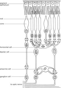
The light-sensitive membrane that lines the interior of the eye. The retina consists of two layers. The inner layer contains nerve cells, blood vessels, and two types of light-sensitive cells (rods and cones). The outer layer is pigmented, which prevents the back reflection of light and consequent decrease in visual acuity. Light passing through the lens stimulates individual rods and cones, which generates nerve impulses that are transmitted through the optic nerve to the brain, where the visual image is formed.
The light-sensitive membrane that lines the interior of the eye. The retina consists of two layers. The outer layer (pigment epithelium) is pigmented, which prevents the back reflection of light and consequent decrease in visual acuity. The inner layer contains nerve cells, blood vessels, and two types of light-sensitive cells (rods and cones). Light passing through the lens stimulates individual rods and cones, which generates nerve impulses that are transmitted through bipolar and ganglion cells to the optic nerve, and hence to the brain, where the visual image is formed. Information can also be transferred horizontally within the retina via a network of horizontal and amacrine cells (see illustration). The retinal neurons are supported by glia called Müller cells.

Structure of the retina
http://webvision.med.utah.edu/book/part-i-foundations/simple-anatomy-of-the-retina/ Anatomy of the retina, prepared by members of the John Moran Eye Center, University of Utah
- edge triggering
- Edgeworth box
- Edgeworth expansion
- Edgeworth, Francis Ysidro (1845–1926)
- Edgeworth price index
- Edgeworth–Kuiper Belt
- EDI
- Ediacaran
- Ediacaran fauna
- Ediacaran fossils
- Ediacaran period
- EDIFACT
- EDI service provider
- Edison cell
- Edison, Thomas Alva
- edit
- edit detection
- editor
- editor software
- editor war
- EDI translator
- Edmonds’ algorithm
- Edmund I (921–46)
- Edmund II (993–1016)
- Edo, Treaty of (1858)