The use of ultrasonics to form an image or representation of an object (such as an internal part of the human body). Ultrasonic tomography is used for these purposes.
In ultrasonic imaging, a probe containing a piezoelectric crystal acts both as the source and the detector of the ultrasonic waves, which are typically in a frequency range of 1–15 MHz. Pulses of ultrasound are transmitted into the body and are reflected at boundaries between different tissue types. By analysing the time lapse and absorption of the returning signals, positions and orientations of the underlying organs and tissues may be deduced.
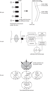
The accompanying diagrams describe the main features of three main types of ultrasound scan employed in modern medicine. In the M-scan, ultrasound is transmitted into the patient’s body. The delay is used to calculate the depth of the structure under examination. Electronics converts the received signal into a potential difference, which is fed to the X plates of a cathode-ray oscilloscope. The timebase of the oscilloscope becomes the time axis of the M-scan. Regular oscillations of the structure under the skin produces a wavy trace. The M-scan techniques are often used for monitoring heart movement. However, the viewing of the heart is limited by surrounding bone and air. Care has to be taken to avoid the ribs and lungs. These restrictions mean that ultrasound cardiography (UCG) requires considerable skill.
Ultrasonic imaging.
In the A-scan, the surfaces in the patient’s body produce echoes, which are picked up by the probe and analysed. A block diagram of the electronics that finally produces the A-scan is shown on the right. The A-scan is a range-finding scan. The surfaces a, b, c, and d produce echoes of the detected ultrasound. The depth of the surfaces are effectively shown by the time delay on the circular screen. The height of a spike which corresponds to a surface represents the size of the echo. The rate generator triggers the transmitter of ultrasound, the swept-gain amplifier, and the timebase simultaneously. The function of the swept-gain amplifier is to increase the return pulse amplification, since echoes returning from deeper structures will be more attenuated. The swept-gain amplifier therefore amplifies the received signals just enough to compensate for the attenuation. The output of the receiver is fed into the cathode-ray oscilloscope which displays the A-scan. A-scans are used when precise depth measurements are required, e.g. in echoencephalography, the determination of the midline of the brain.
In the B-scan, the ultrasound probe is moved slowly over the patient’s body and a cathode-ray oscilloscope is able to store the echoes of a succession of range-finding scans displays them as bright spots. The depth is calculated by the delay of the return signal and the brightness of the spots indicates the strength of the return signal. The two-dimensional images produced by B-scans are ideal for monitoring the progress of a foetus in a womb or locating tumours or other anomalies in the liver, kidneys, and ovaries. B-scans are also invaluable in the examination of aneurysms (swollen parts of arteries).
Ultrasound can also be used to monitor the function of organs or blood flow. This application of ultrasound relies on the Doppler effect. The transmitter is separate from the receiver. The ultrasound used in this technique is a continuous beam rather than the series of pulses used for imaging. The transmitted and received signals are electronically mixed and the output is filtered so that only the Doppler-shift frequency is amplified. Doppler-shift frequencies are often in the human hearing range so that the operators can ‘hear’ the moving structures on earphones.
Ultrasound monitoring using the Doppler effect is also used in monitoring blood flow. The Doppler probe is placed on the skin directly above a blood vessel at an angle θ to the horizontal. The blood, travelling in a direction parallel to the skin, reflects the transmitted waves and Doppler-shifts them by an amount Δf given by the formula:
where f is the frequency of the transmitted ultrasound, ν is the speed of the blood flow, and c is the speed of sound in blood. A blockage due to a clot (thrombosis), or a constriction due to plaque build-up on the vessel walls (atheroma), is immediately apparent if there is a change in the Doppler shift frequency.
Ultrasound may also be delivered at large intensities to localized regions of the body for therapeutic purposes. Diathermy is the application of ultrasound to cause localized heating of tissue to relieve pain in the joints of arthritis sufferers. Typical treatments use intensities of several W/cm2 for periods of a few minutes several times a week. Actual destruction of tissue is also possible using sufficiently high intensities of ultrasound (intensities exceeding 1000 W/cm2). Ultrasound has been shown to destroy cancer cells, although some studies have also shown that it can in some cases stimulate growth of the tumour.